

More:
News
Paper written by two students from the Faculty of Engineering published in Applied Sciences magazine
The students, Alex Liebenson and Mark Rayhshtat, guided by Dr. Dror Malka, based their paper on their Final Project and it deals with improving the efficiency of the diagnosis of MS using MRI scans.
"In this paper, we present a new method of displaying Magnetic Resonance (MR) images taken from Multiple Sclerosis (MS) patients. We show that our method can potentially make the diagnostic process far more focused and concise. The method is implemented as an algorithm-based application, which automatically detects MS lesions and reduces the amount of reviewed images by 98% or more. In contrast to existing detection algorithms, our application utilizes five different types of MR images as well as the Digital Imaging and Communications in Medicine (DICOM) standard, supporting a wide range of data sets. After images are selected for file type and relevant brain region, each image is subjected to four separate algorithms, the results of which are combined into a single displayed image for the use of the diagnosing physician.
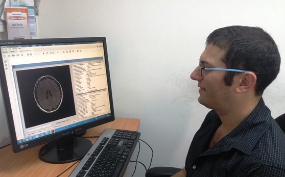
Current MR consists of 500–700 images of various types for each tested subject; as m
any as thirty for a single type (accounting for the different coordinates of the virtual brain slices). Additionally, within images that belong to a suitable type, a significant portion is concerned with brain areas that are irrelevant for the diagnosis (i.e., images of areas without WM lesions). Altogether, the vast majority of images are not useful for diagnostic purposes. Currently, decoding of these images is performed manually by a specialized diagnostic physician who reviews each image separately, and looks for images which are relevant for the identification of MS lesions, if any exist. The problem we address is that manually reviewing a large collection of images makes the process lengthy and complicated. In this study, we present an automated solution to the problem, a solution that consists of several algorithms including file type selection, image enhancement and lesion detection. The combination of these algorithms is used to reduce the amount of relevant images presented to the diagnosing physicians by more than 98% while enhancing the information in each presented image, therefore greatly improving the efficiency of MS diagnostic process".
- News & Events
